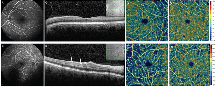
Figure 2.

Fundus fluorescein angiography shows complete perfusion of the retinal vessels in both eyes (A, B). Spectral domain optical coherence tomography (SD-OCT) scan of the left eye shows inner nuclear layer thinning associated with outer plexiform layer elevation consistent with resolved paracentral acute middle maculopathy (PAMM) (C). SD-OCT scan of the right eye reveals a hyperreflective parafoveal band at the level of the inner nuclear and inner plexiform layers corresponding to acute PAMM (D). OCT angiography shows normal perfusion of the deep and superficial capillary plexuses in the left eye (E, G) and decreased perfusion of the deep capillary plexus but normal perfusion of the superficial capillary plexus in the right eye (F, H)
