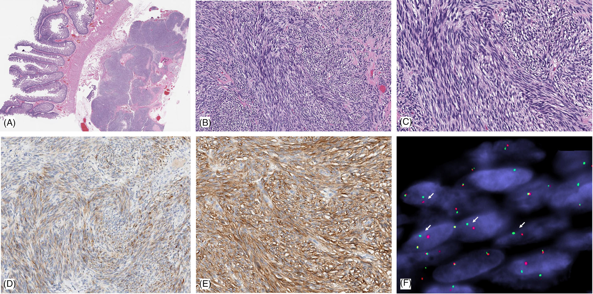
FIGURE 1.

Pathologic features of case 1: small intestinal GIST harboring a novel AGAP3-BRAF fusion. Low power view shows a lobulated tumor within the muscularis propria and subserosal layer of the small bowel (A). Intermediate power showing highly cellular, intersecting fascicles of spindle cells (B); while at high power show eosinophilic cytoplasm, intracytoplasmic vacuoles, and monomorphic nuclei with fine chromatin. (C). Immunohistochemically the tumor showed weak staining for KIT/CD117 (D), while there was diffuse, strong staining for DOG1 (E). (F). FISH shows break-apart red (centromeric) and green (telomeric) in keeping with a BRAF gene rearrangement (the narrow, fixed gaps between the break-apart signals support an intrachromosomal inversion)
