
FIGURE 2.
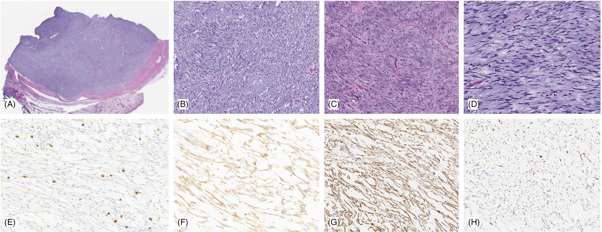
Pathologic features of distal esophageal GIST with MKRN1-BRAF fusion. Low power shows a well-circumscribed lesion surrounded by a fibrous capsule (A), which is composed of loose fascicles of bland spindle cells with scant eosinophilic cytoplasm and ovoid unform nuclei with fine chromatin (B,C). The tumor is associated with extensive myxoid stroma and scattered mast cells (D). Immunohistochemically the tumor cells were negative for KIT/CD117 (E) (which highlights the stromal mast cells, as internal positive control), while diffusely positive for DOG1 (F) and SMA (G), and only rare cells label with desmin (H)