. 2021 Nov 1;10:e72132. doi: 10.7554/eLife.72132

Figure 5. Model simulations recapitulate experimentally observed phenotypes through genetic, pharmacological, and mechanical perturbations.

(A) Reference model simulation of the wild-type scenario. The figure displays a schematic representation of the auxin flow inside the root (left picture), cell growth rate (right picture). The bottom graph shows the profiles of auxin concentration in the vascular tissues (dashed blue line), auxin concentration in the non-vascular tissues (dotted blue line,) and growth rate (red line) along the root axis. (B) Model simulation of the pin2 knockdown mutant. In silico pin2 mutant shows strongly reduced PINs expression in the lateral root cap, epiderm, is, and cortex. Note that acropetal auxin flow is severely affected and auxin tends to accumulate in the lateral tissues as observed in experiments (Dhonukshe et al., 2010). (C) Mechanical removal of lateral root cap resulted in the strong accumulation of auxin inside the root tip, largely because auxin cannot flow anymore shootward through outermost tissues whereas growth rate was not significantly affected. (D) Simulation of root tip cutting. Removing the root tip results in a general increase of auxin level in the central vascular tissues, as a consequence of the disappearance of acropetal auxin flow. PINs localization in the external tissues is also affected due to the loss of incoming auxin flow. (E) Simulated CMTs disruption (i.e. oryzalin treatment or similar) on root growth and polarity. CMTs disruption was simulated by inducing a fast degradation of the anisotropy factor. Cells lose polarity and growth anisotropy, causing the root to expand and bulge radially as observed in experiments (Baskin et al., 1994). Notice that the top cell row is considered to be a static attachment of the root to the substrate and therefore its growth is not affected during the simulation. (F) Legend and scale bars of auxin concentration and cell growth rate. ‘Auxin conc. Vasc.’ indicates auxin concentration in the vascular central tissues (the vascular cells and the pericycle), while ‘Auxin conc. non-Vasc.’ indicates auxin concentration in the remaining external tissues and the root tip. The simulations have been run for 1500 time steps.

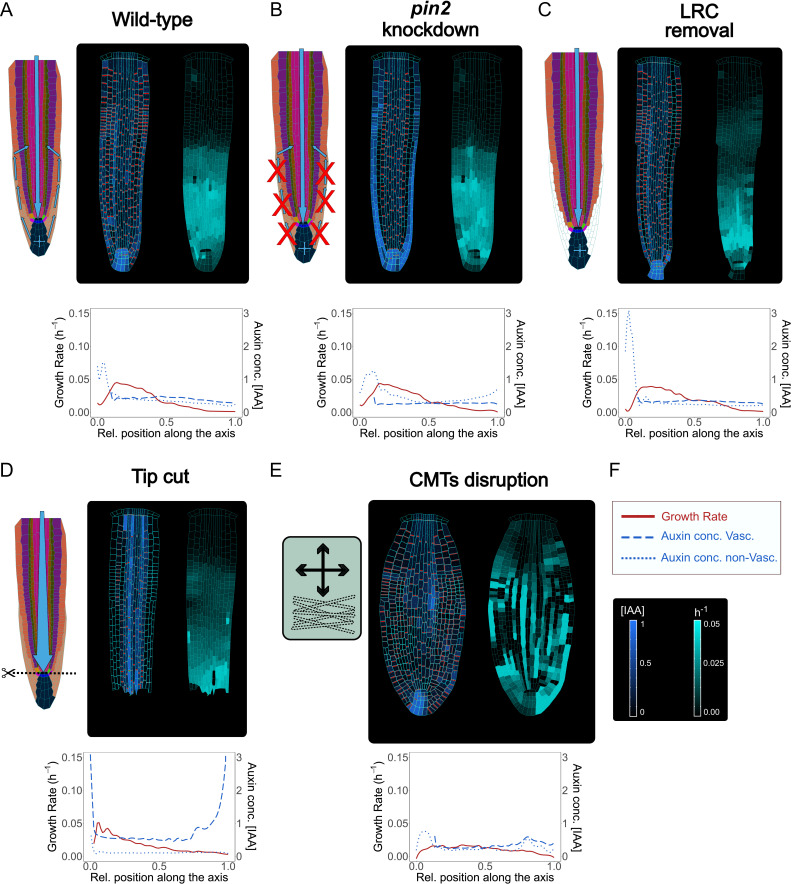
Figure 5—source data 1. Source data used to generate Figure 5A-E.

Figure 5.

Figure 5—figure supplement 1. Model simulations recapitulate some of the experimentally observed phenotypes, related to Figure 5.

Figure 5—figure supplement 1.

(A) Model simulations of the vascular PINs reduction. PINs expression was reduced in the vascular tissues, pericycle, and endodermis, to maintain 10 % PIN activity. The model predicts that auxin levels in the root are strongly depleted as the hormone flow is severely affected by PIN down-regulation. (B) Simulated aux1 knockdown mutant. AUX/LAX expression was reduced by 90 % in every cell. This simulated mutant present decreased auxin levels and root growth. (C) Model simulations of QC ablation. Removing the QC cells results in an increase of auxin level in the vascular initial cells above the ablated QC. The model simulations display the disappearance of acropetal auxin flow, as much less auxin each and accumulate in the root tip. (D) Legend and scale bars of auxin concentration and cell growth rate. ‘Auxin conc. Vasc.’ indicated auxin concentration in the vascular central tissues (the vascular cells and the pericycle), while ‘Auxin conc. non-Vasc.’ indicates auxin concentration in the remaining external tissues and the root tip. All simulations have been run for 1500 time steps.
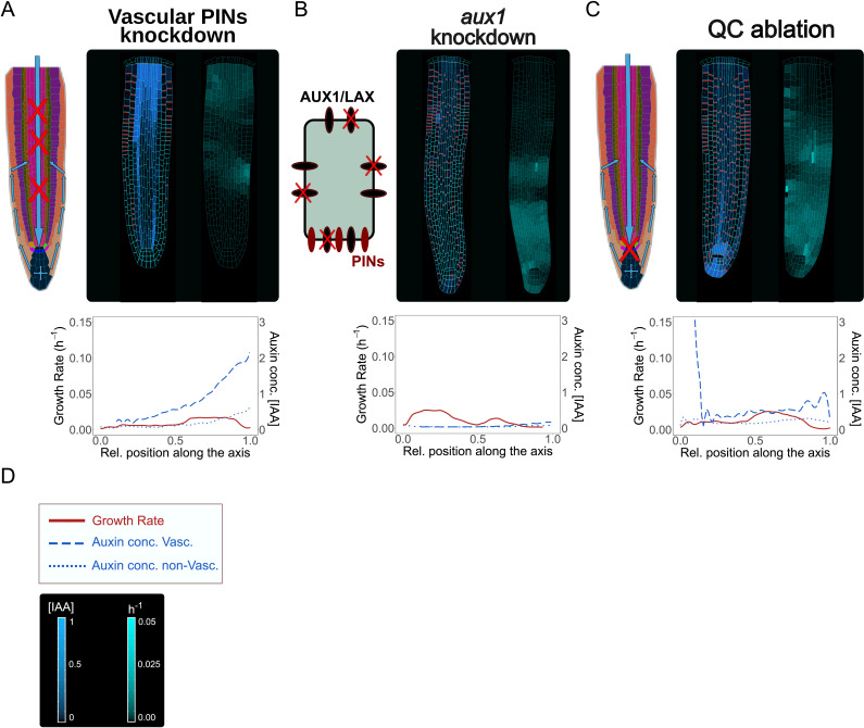
Figure 5—figure supplement 1—source data 1. Source data used to generate Figure 5—figure supplement 1A–1C.
Figure 5—figure supplement 2. Model simulations using alternative wild-type embryo templates.

Figure 5—figure supplement 2.

(A, B) Embryo templates extracted from the following studies Nieuwland et al., 2016 (A); Scheres et al., 1994 (B). The particular choice of an initial embryo template has no impact on the emergence of root growth and auxin distribution. The simulations have been run for 1500 time steps.
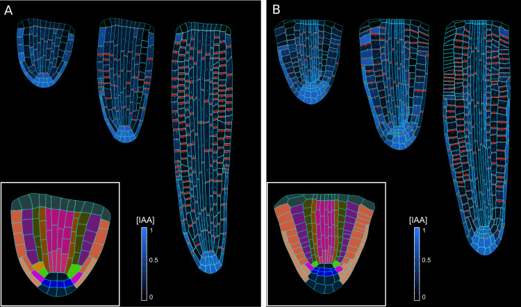
Figure 5—figure supplement 3. Model parameters sensitivity.

Figure 5—figure supplement 3.

(A) Model testing for parameter kP, the coefficient of contribution for auxin flow to PIN sensitivity (Equation. 14). The values tested are (from the left to right): 0, 1, 3 (default wild-type), 4, and 5. (B) Model testing for parameter K1auxin, the auxin-induced cell wall relaxation coefficient (Equation. 27). The values tested are (from the left to right): 0.005, 0.01, 0.05 (default wild-type), 0.1 and 0.2. The simulations have been run for 1500 time steps.
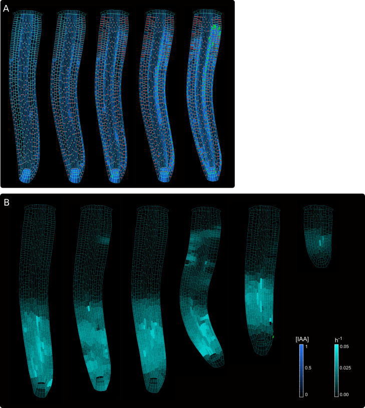
Figure 5—video 1. Model simulations of the pin2 knockdown mutant, related to Figure 5B.
Download video file (993.6KB, mp4)
In silico pin2 mutant shows strongly reduced PINs expression in the lateral root cap, epidermis, and cortex.
Figure 5—video 2. Model simulations of the vascular PINs reduction, related to Figure 5—figure supplement 1A.
Download video file (1MB, mp4)
Figure 5—video 3. Model simulations of the aux1 knockdown mutant, related to Figure 5—figure supplement 1B.
Download video file (1,011.7KB, mp4)
AUX/LAX expression was reduced by 90 % in each cell.
Figure 5—video 4. Model simulation of QC ablation, related to Figure 5—figure supplement 1C.
Download video file (1.2MB, mp4)
Removing the QC cells results in an increase of auxin level in the vascular initial cells just above the ablated QC.
Figure 5—video 5. Model simulation of lateral root cap ablation, related to Figure 5C.
Download video file (1.1MB, mp4)
Mechanical removal of LRC resulted in the strong accumulation of auxin inside the root tip, largely because auxin cannot flow anymore shootward through outermost tissues whereas growth rate was not significantly affected.
Figure 5—video 6. Model simulations of root tip cutting, related to Figure 5D.
Download video file (1,012.2KB, mp4)
Figure 5—video 7. Model simulation of mechanics disruption on root growth and polarity, related to Figure 5E.
Download video file (1.2MB, mp4)