. 2021 Dec 29;10:e71981. doi: 10.7554/eLife.71981

Figure 4. Dopaminergic (DA) axons make rare synapses on the soma and shaft of nucleus accumbens (NAc) resident neurons.

Three examples of DA axons making synapses on the soma (left column) and shaft (right column) are depicted. Mito-Apex2 DA axons are shaded in light blue with an asterisk marking an Apex2-positive mitochondria which are significantly darker than Apex2-negative mitochondria (green arrowhead). Red arrows point to the post-synaptic density (PSD) formed between the DA axon and soma. Pie chart in upper left corner shows ratio of synaptic to non-synaptic DA axon varicosities. Scale bar = 500 nm.

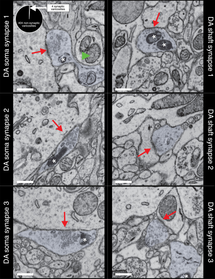
Figure 4.

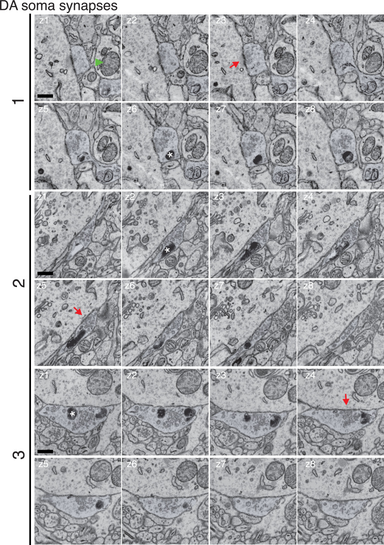
Figure 4—figure supplement 1. Montage of identified dopaminergic (DA) axons making soma synapses.

Figure 4—figure supplement 1.

Three (1–3) putative DA soma synapses shown in Figure 4 are more clearly depicted by showing eight serial electron microscopy (EM) sections spanning the synapse (z1–z8). Red arrows point to the post-synaptic density (PSD), green arrowhead shows Apex2-negative mitochondria for comparison to Apex2+ mitochondria, marked with an asterisk. Scale bar = 0.5 µm.

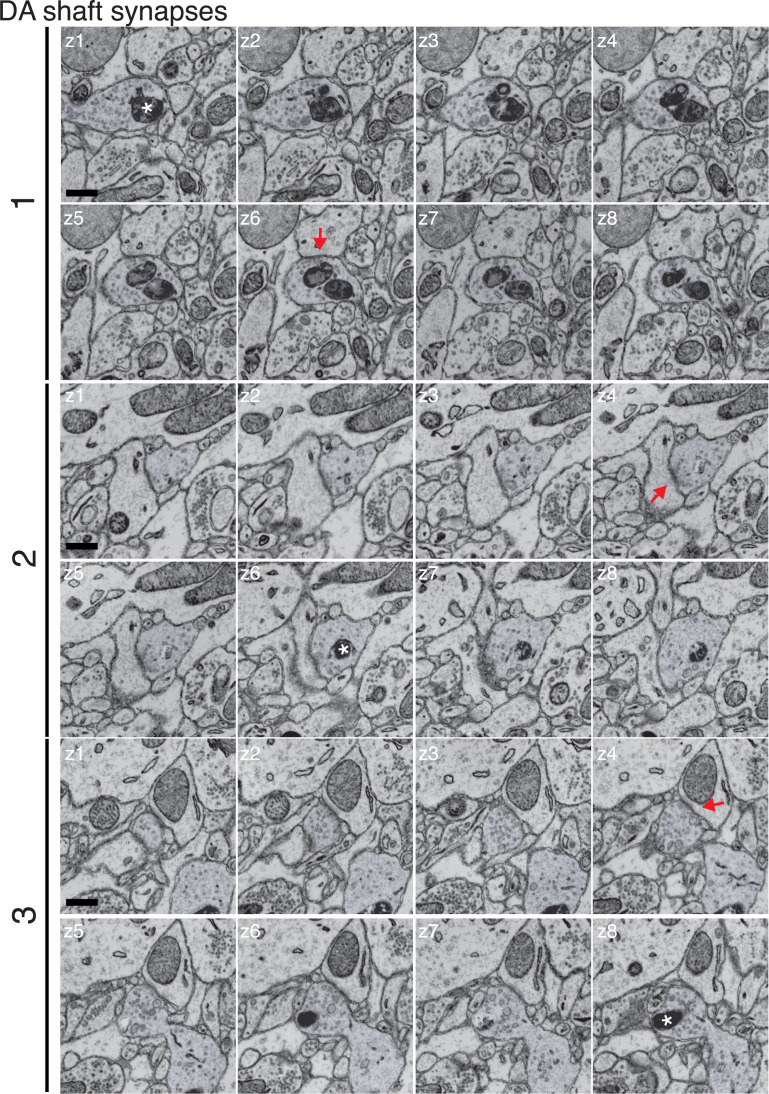
Figure 4—figure supplement 2. Montage of identified dopaminergic (DA) axons making shaft synapses.

Figure 4—figure supplement 2.

Three (1–3) putative DA shaft synapses shown in Figure 4 are more clearly depicted by showing eight serial electron microscopy (EM) sections spanning the synapse (z1–z8). Red arrows point to the post-synaptic density (PSD), asterisks marks the Apex2+ mitochondria. Scale bar = 0.5 µm.

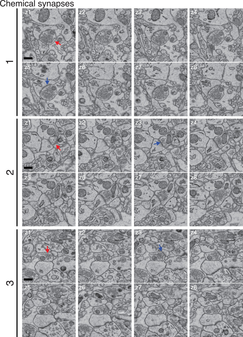
Figure 4—figure supplement 3. Montage of representative chemical synapses.

Figure 4—figure supplement 3.

Three (1–3) chemical synapses identified by a prominent vesicle cloud and post-synaptic density (PSD) in apposition to a post-synaptic neurite were identified in the 6 nm resolution mito-Apex2 dataset. For each example, eight serial electron microscopy (EM) sections spanning the synapse were placed in order (z1–z8). Red arrows point to vesicle cloud and blue arrows point to the PSD. Scale bar = 0.5 µm.