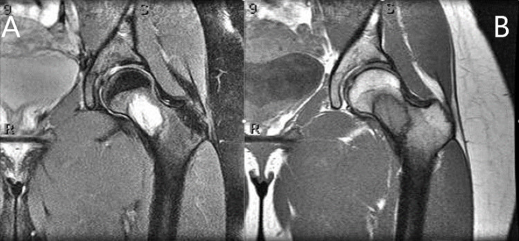
Figure 2. MRI scan of the left hip.
MRI scan of the left hip with T2-weighted coronal image (A) demonstrating a hyperintense isolated lesion with distinct sclerotic margins and surrounding bone marrow edema within the femoral neck and a T1-weighted coronal image (B) showing an isointense lesion with sclerotic margins

