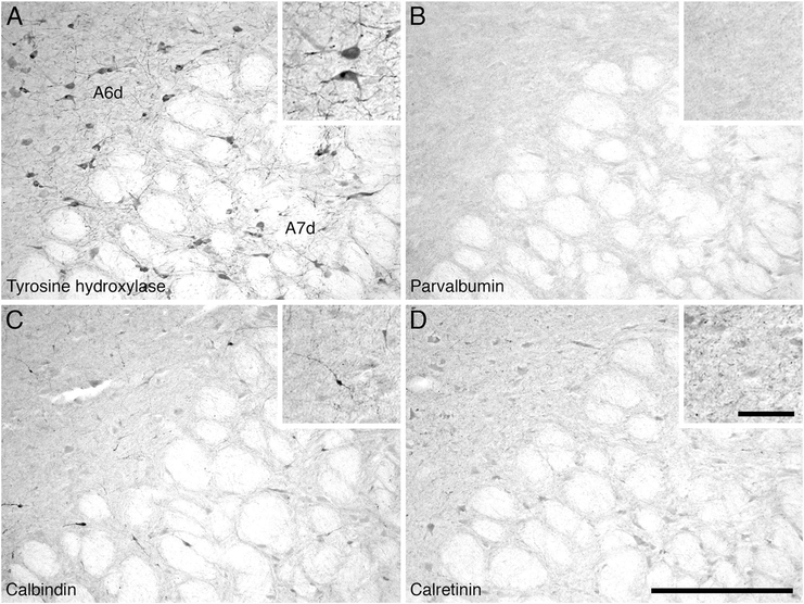
Figure 12.
Low (main image) and high (inset) magnification photomicrographs in the region of the diffuse division of the locus coeruleus (A6d) and the subcoeruleus diffuse division (A7d) of the locus coeruleus complex in the pons of the river hippopotamus. A: Neurons immunoreactive for tyrosine hydroxylase showing the diffuse portions of both the locus coeruleus (A6d) and subcoeruleus (A7d). Note the absence of structures immunoreactive for parvalbumin (B), the moderate density of calbindin structures (C), and the very low density of structures immunoreactive for calretinin (D) in these nuclei. In all images medial is to the left and dorsal to the top. Scale bar = 500 μm in D (applies to A–D); 100 μm in inset to D (applies to all insets).

