
FIGURE 1. Extracellular vesicles (EVs) isolated from burnmice induce cytokine dysfunction upon adoptive transfer to uninjured mice in vivo.
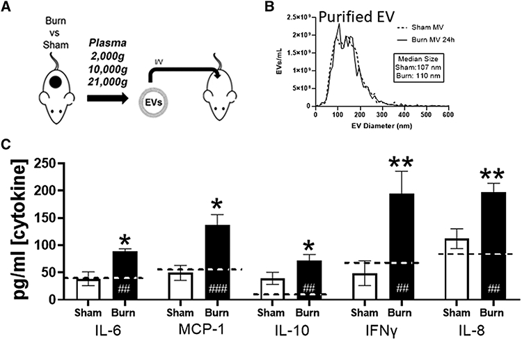
(A) Experimental design. Mice underwent 20% total body surface area (TBSA) burn injury or sham injury and EVs were isolated from plasma (n = 12 mice per group) 48 h after injury. EVs were pooled from sham-injured (white bars) and burn-injured (black bars) mice (n = 6 per pool) were adoptively transferred into uninjured healthy mice (n = 3/group per experimental replicate) at a concentration of ~12.5% of baseline total circulating plasma EVs (1010/mouse) i.v. by tail vein injection. Recipientmice were sacrificed 24 h after EV transfer. (B) Nanoparticle tracking analysis (NTA) analysis was to use to measure frequency and size distribution (i.e., diameter) of EVs isolated from burned and sham mice prior to transfer. No differences in size distributions of isolated EVs were found after burn or sham treatment. (B–G) Plasma concentrations of free cytokines after cell stimulation by burn EV (solid bars), sham EV (open bars), or untreated mice (dashed line) were measured by Bio-Plexmultiplex analysis. This experiment was performed in triplicate and data pooled, where *P < 0.05 and **P < 0.005 for burn EV-treated mice vs. sham EV-treated mice; ##P < 0.005 and ###P < 0.001 EV treated mice vs. normal, untreated mouse plasma levels of cytokine; n = 6/group *P < 0.05, t-tests with FDR q = 0.03