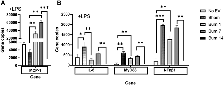
FIGURE 9. Extracellular vesicles (EVs) isolated from key time points after burn injury induce significantly different levels of LPS-responsive innate immune genes inmacrophages in vitro.

mRNA was isolated from RAW264.7 cells stimulated with 1 μg/ml LPS for 24 h in the presence or absence of 3 × 10⁵ EVs isolated from the plasma 1, 7, and 14 d after burn injury (each group represents cell cultures stimulated with EVs from 12 source mice, with n = 6 different pooled EV preparations from 2 individual mice). Gene expression was evaluated using Nanostring barcoding spanning 561 mRNAs (nCounter Mouse Immunology CodeSet v3.0). Data are presented as the gene copy numbers of MCP-1, IL-6, MyD88, and NFkB1 after data normalization to housekeeping and internal control genes by nSolver v4.0. Data shown ± SEM; *P < 0.05, **P < 0.01, ***P < 0.005 by Mann-Whitney unpaired nonparametric t-test
