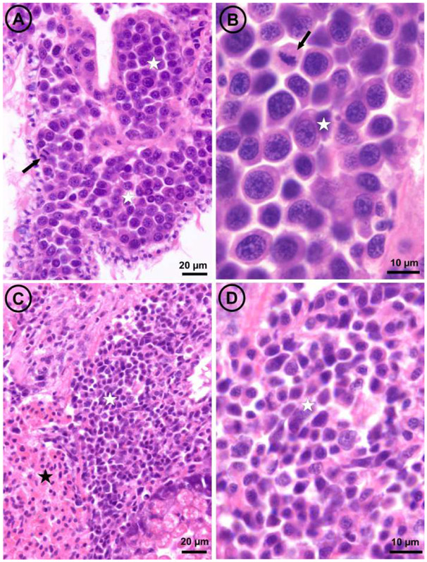
Fig. 4: Light micrographs of histological sections through the visceral mass of two mussels,
one affected by disseminated neoplasia type A (panels A and B) and the other by disseminated neoplasia type B (panels C and D). The arrows point out mitotic figures; white stars mark masses of neoplastic cells; the black star marks a mass of normal hemocytes.

