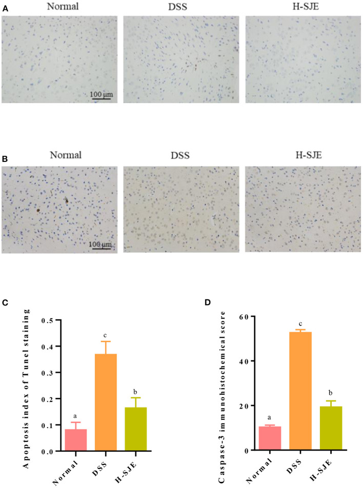
Figure 6.
Effects of SJE on cerebral apoptosis. (A) Representative photomicrographs, demonstrating the detection of apoptotic cells by TUNEL stain (200X). (B) Representative immunohistochemical photomicrograph of Caspase-3 (200X). (C) Analysis of apoptosis index and (D) immunohistochemical score of Caspase-3 score. Data are presented as mean ± SEM, n = 5. All statistical tests were conducted using one-way analysis of variance (post-hoc test: Duncan) and values designated by different letters were statistically different (p < 0.05).

