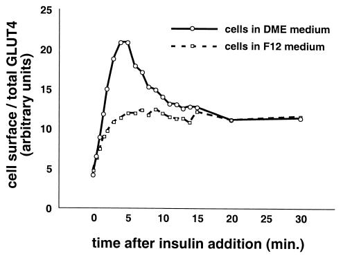
FIG. 5.
Culture conditions modulate the kinetics of insulin-stimulated GLUT4 translocation in CHO cells. Two days before the experiment, confluent CHO cells were placed in DMEM identical to that used for 3T3-L1 adipocytes or left in F12 culture medium. Cells were starved overnight before the experiment. On the day of the experiment, cells were stimulated with 80 nM insulin for 0, 0.5, 1, 1.5, 2, 3, 4, 5, 6, 7, 8, 9, 10, 11, 12, 13, 14, 15, 20, or 30 min, then transferred to 4°C, and washed with cold PBS. Staining and flow cytometry were done as described in the text. For cells cultured in DMEM, insulin stimulated a rapid increase in GLUT4 reporter present at the cell surface, peaking at 4 to 5 min after insulin addition. Subsequently, the proportion of GLUT4 on the plasma membrane decreases, and a steady state in the presence of insulin is reached 20 min after insulin addition. In contrast, cells cultured in F12 medium externalized GLUT4 with monophasic kinetics, characterized by no overshoot of the final steady-state response in the presence of insulin. Both the initial and final proportions of GLUT4 at the cell surface are essentially unaffected by the culture conditions, despite the marked effect on intermediate time points. For cells cultured in DMEM, the peak response is a 5.5-fold increase in the proportion of GLUT4 at the cell surface compared to the basal state. The kinetics and the amplitude of the peak response are similar in CHO cells cultured in DMEM and in 3T3-L1 cells (compare to Fig. 3a, 4a, and 4c).

