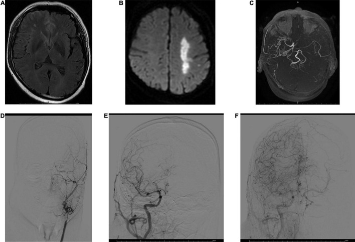
FIGURE 2.
MRI and digital subtraction angiography (DSA) images obtained 3 h after stroke onset in a 53-year-old male. FVHs were observed at the left temporooccipital junction (A). DWI shows acute infarction in the territory of the left MCA (B). FVHs located beyond the DWI lesions, indicating an FVH–DWI mismatch. Magnetic resonance angiography (MRA) (C) shows near-occlusion of the left MCA and ICA. A DSA image obtained soon afterward demonstrates stenosis of the left common carotid artery and occlusion of the ICA (D). Angiograms of the right common carotid artery in anteroposterior view in the early arterial phase (E) and late arterial phase (F) show good retrograde filling of the leptomeningeal collateral circulation in the left hemisphere.

