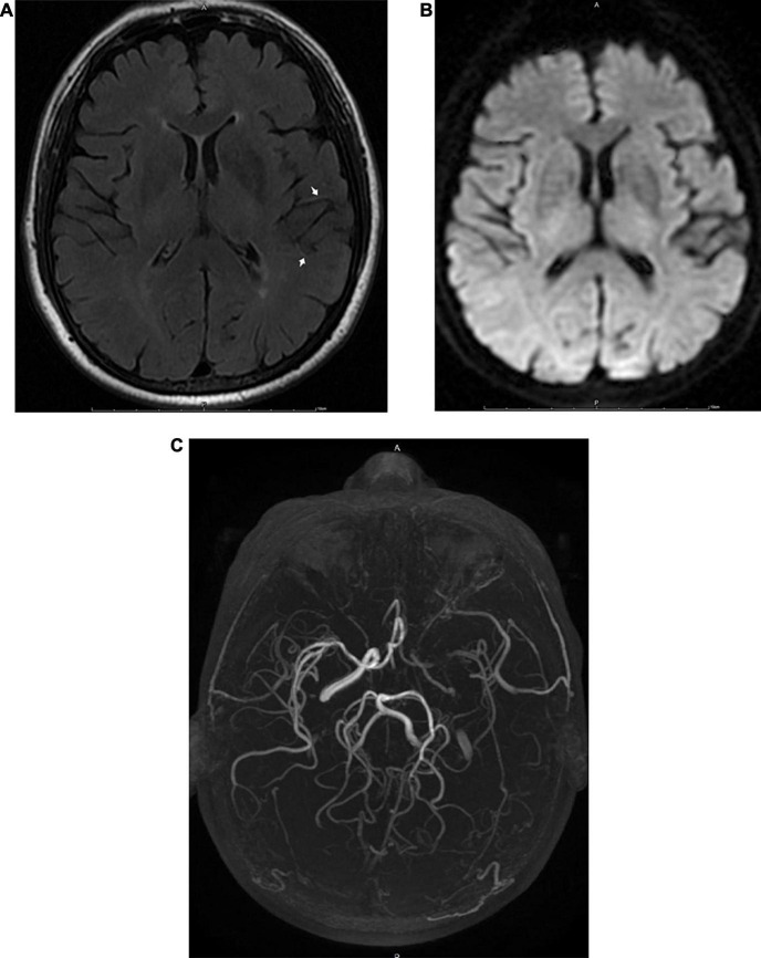
FIGURE 3.
MRI images obtained 6 h after the appearance of the initial symptoms in a 68-year-old male patient with transient ischemic attack. A FLAIR image (A) shows FVH in the territory of the left MCA (arrow). There were no abnormalities on DWI (B). Three-dimensional time-of-flight MRA (C) shows occlusion of the ipsilateral MCA and ICA.

