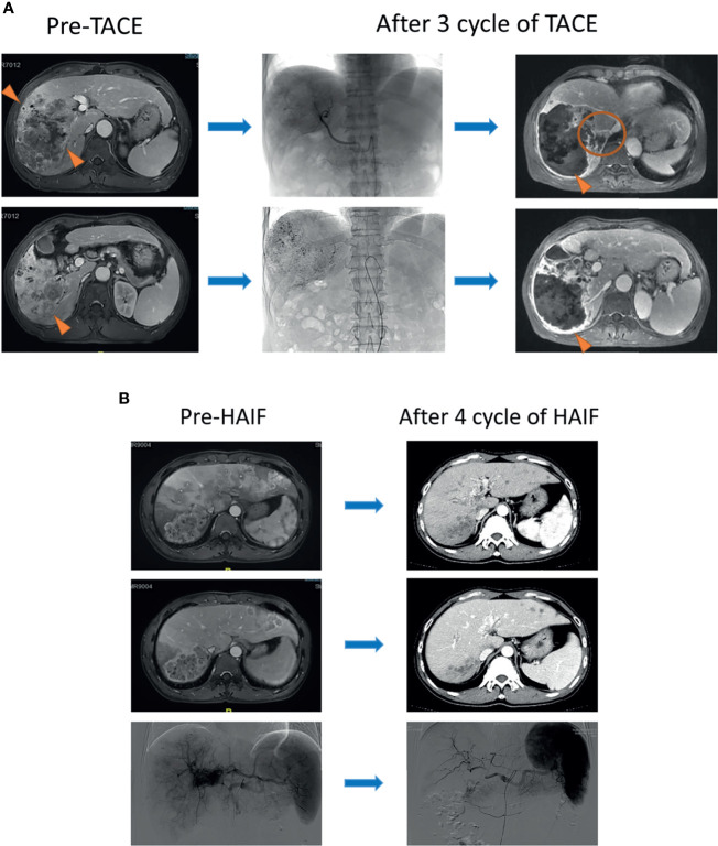
Figure 2.
(A) A 57-year-old male patient was diagnosed with infiltrative HCC by contrast MRI scanning and received three-cycle TACE. Obvious necrosis appeared in the center of the tumor after therapy (triangle), but the marginal lesions became enlarged homogeneously (circle). (B) A 46-year-old male patient with multiple lesions was diagnosed with infiltrative HCC. Portal vein fistula was revealed at the arterial phase of the MRI scanning image. After a four-cycle HAIC, all of the lesions in the liver apparently shrank (triangle).

