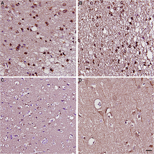
FIGURE 9.

Immunohistochemical staining of CARMIL3 in human infarct brains and stroke-free autopsied brains. (A–C) CARMIL3 expression in human infarct brain tissues. In the region most sever ely infarcted (right lower part of A), there were fewer neurons, with all the residual neurons strongly positive for CARMIL3. In the area surrounding the sever ely infarcted region, most neurons had strong CARMIL3 staining in their nuclei and cytoplasm. In the region with the least infarction severity (left upper part of A), most neurons had normal nuclear morphology with faint CARMIL3 staining mainly in the cytoplasm. (D) Relatively low CARMIL3 expression in the neurons of stroke-free autopsied brains. Scale bar = 20 μm.
