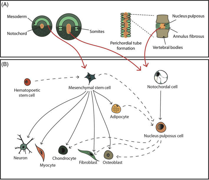
FIGURE 1.

Cell sources and linages involved in cellular therapy in regenerating the intervertebral disc. (A) Schematic illustration depicting key stages of intervertebral disc development, highlighting the mesodermal origin of the notochord and sclerotome that evolve into the nucleus pulposus, annulus fibrosus and vertebral bodies. Red arrows show the potential cell sources. (B) An illustration of the mesenchymal stem cell and notochordal cell differentiation lineages (black arrows). Under appropriate culture conditions transdifferentiation can be induced to develop different cell types (black dash arrows), which can interlink different cell lines
