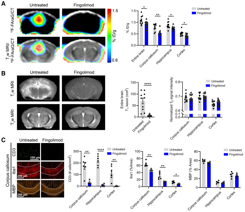
FIGURE 6.
(A) 18F-FAraG PET/CT and 18F-FAraG PET/CT overlaid on T2-weighted (T2w) MR images from untreated and fingolimod-treated animals at W7–14dpi. Graph shows quantification of 18F-FAraG signal in entire brain, corpus callosum, hippocampus, and cortex. (B) T1-weighted (T1w) and T2-weighted MR images of fingolimod-treated and untreated mice and corresponding quantification of T1-enhancing lesions and normalized T2-weighted signal intensity of corpus callosum, hippocampus and cortex. (C) Immunofluorescence images of corpus callosum for CD3 T cells (green), microglia/macrophages (Iba1, red), and myelin (MBP, orange) from untreated and fingolimod-treated mice at W7–14dpi. Graphs show quantification of immunofluorescence images from corpus callosum, hippocampus, and cortex. ID = injected dose. *P ≤ 0.05. **P ≤ 0.01. ****P ≤ 0.0001.

