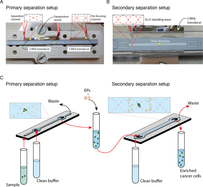
Figure 1.
Overview of the of two-step acoustophoresis (A2). (A) Photograph of the primary separation chip and aluminum chip holder prior to assembly. Showing the prefocusing channel followed by the separation channel, the two piezoelectric transducers for sound generation, as well as a temperature sensor. (B) Photograph of the multinode (3λ/2) purging chip with one piezoelectric transducer and separation channel. (C) Schematic of the workflow and separation principle in A2. In the primary separation setup, a cell sample input represented by white (WBCs) and green (cancer cells) circles enters the chip through the prefocusing channel. After passing through the separation channel, the cells are collected at the central outlet. The cells are incubated with elastomeric particles (EP) and subsequently processed through the secondary multinode separation chip. The purified cancer cell fraction is collected at the central outlet.

