Figure 4:

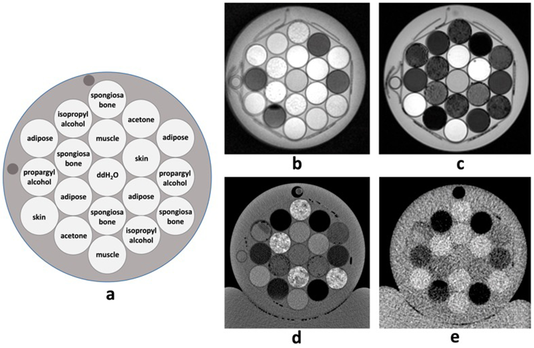
Axial representation of a) example phantom configuration and corresponding images of b) ZTE (1H) proton density-weighted MRI, c) Dixon water-only MRI, d) kVCT, and e) MVCT. Each image depicts a single slice at the same level.

Axial representation of a) example phantom configuration and corresponding images of b) ZTE (1H) proton density-weighted MRI, c) Dixon water-only MRI, d) kVCT, and e) MVCT. Each image depicts a single slice at the same level.