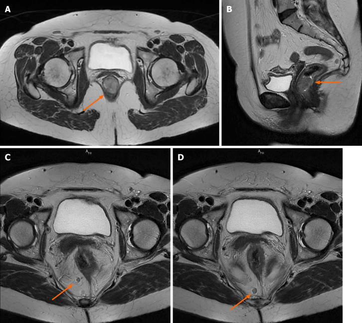
Figure 3.
Pelvic magnetic resonance imaging scan with intravenous gadolinium administration. A and B: Axial and sagittal T2-weighted images showing the tumor occupying the anorectal junction canal, with homogenous enhancement, without infiltration of the rectal fascia; C and D: Axial T2-weighted propeller images showing two large lymph nodes located in primary stations, without mesorectal fascia retraction.

