Figure 1.

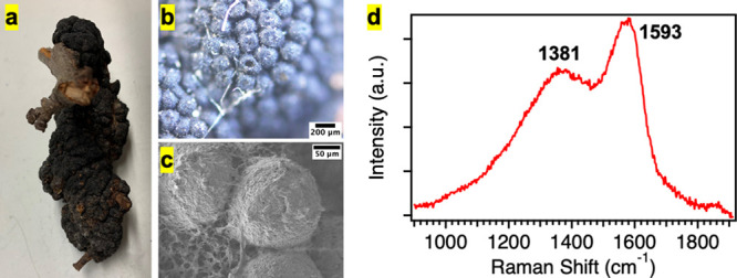
(a) Picture of a black knot (A. morbosa). (b, c) Optical and SEM images of the black knot fungus, where the knot appears as a cluster of ∼150 μm nodules. Scale bars are indicated on the images. (d) Representative Raman spectrum of the black knot showing characteristic melanin peaks at 1381 and 1593 cm–1.
