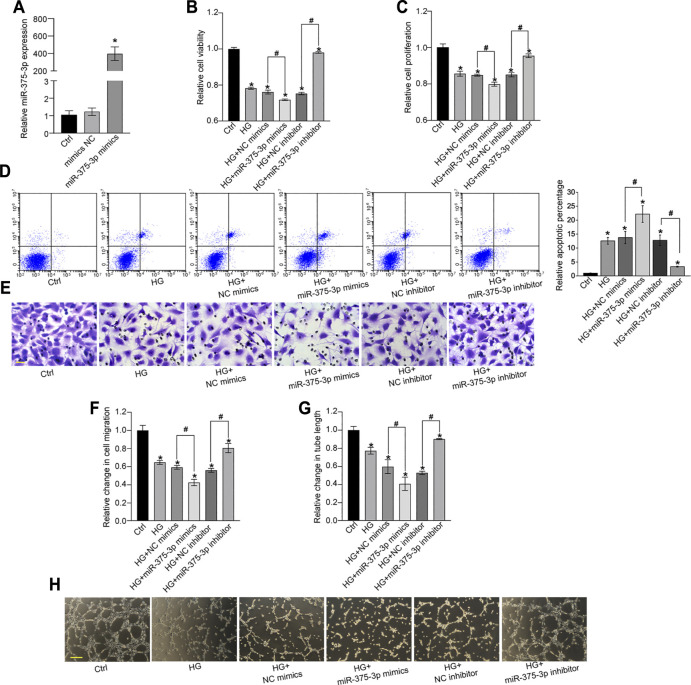
FIGURE 4.
miR-375–3p regulates EPC function in vitro. (A) EPCs were transfected with negative control (NC) mimics, miR-375–3p mimics, or left untreated (Ctrl) for 24 h. The expression of miR-375–3p was detected by quantitative reverse transcription-polymerase chain reaction (qRT-PCR) (A). (B–H) EPCs were transfected with NC mimics, miR-375–3p mimics, NC inhibitor, miR-375–3p inhibitor, or left untreated (Ctrl) for 24 h and then treated with or without high glucose (30 mM) for 24 h. The viability of EPCs was determined by MTT assays (B). The proliferation ability of EPCs was determined by CCK-8 assays (C). Flow cytometry assays and quantitative analysis were conducted to detect cell apoptosis (D). Cell migration and quantitative analysis was conducted using Transwell assays [(E,F), scale bar: 20 μm]. Tube formation assays and quantitative analysis were conducted to detect the tube-formation activity of EPCs [(G,H), scale bar: 50 μm]. *p < 0.05 versus Ctrl group; Pound sign (#) indicates significant differences between the marked groups. All experiment was repeated three times.

