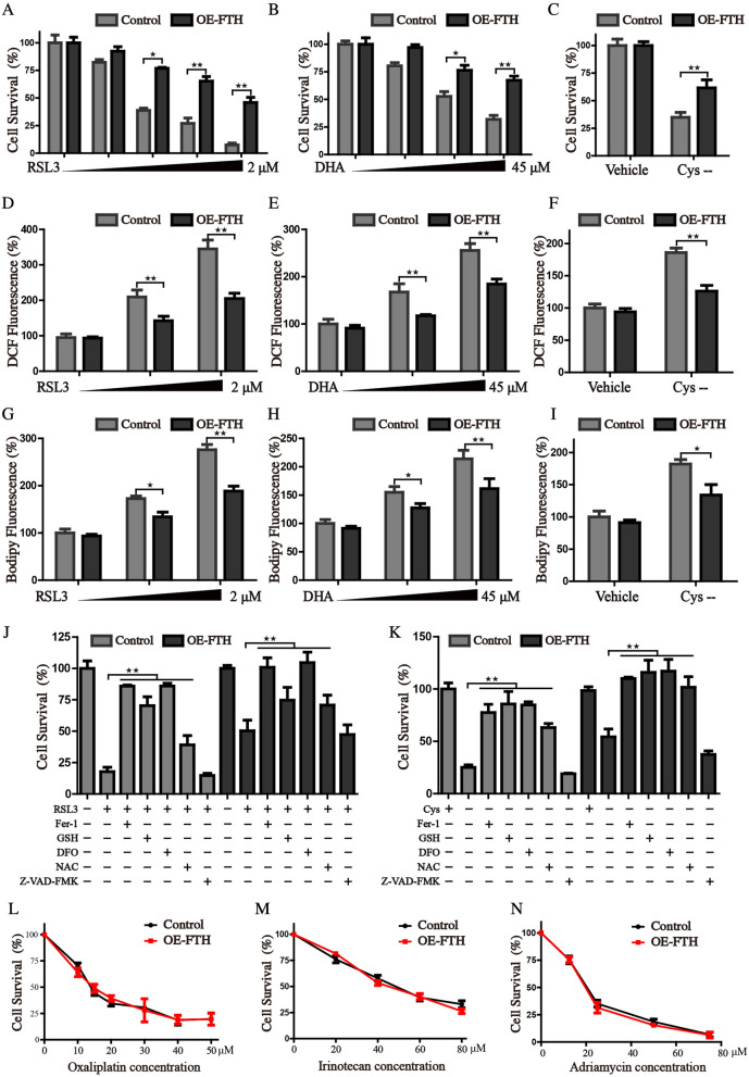
Fig. 4.
FTH inhibits the ferroptosis in HCC cells. A–C Control and FTH overexpressed cells were treated with RSL3 (0–2 μM), DHA (0–45 μM), or cystine-free medium, the cell survival rate was monitored by CCK8 assay. Cellular ROS (D-F) and lipid ROS production G–I were measured by the staining of DCF-DA and Bodipy after indicated treatment. J, K Control and FTH overexpression cells were treated RSL3 or cystine-free medium in the presence or absence of cell death inhibitors. The cell viability was detected by CCK8 assay. L–N FTH enforced expression and control cells were treated with Oxaliplatin (0–50 μm), Irinotecan (0–80 μm), or Adriamycin (0–72 μm) for 36 h, and the cell survival was measured using CCK8 assay. (Values are represented as mean ± SD. ★P < 0.05, ★★P < 0.01 versus indicated groups)

