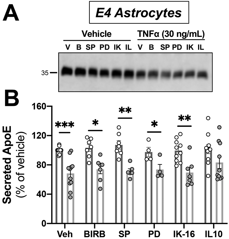
Figure 6. Astrocytic apoE4 secretion reduction after TNFα activation is unaltered across several inflammatory signaling pathways.

Astrocytes from human APOE4 targeted-replacement mice were treated with 30 ng/mL of TNFα and inhibitors. Conditioned media was collected at 24 hrs., and was analyzed by western blotting with antibodies against apoE. Veh = vehicle; BIRB = Doramapimod, p38 MAPK inhibitor (1 μM); SP = SP600125, JNK kinase inhibitor (10 μM); PD = PD98059, MEK inhibitor (50 μM); IK-16 = IKK-16, IkappaB inhibitor (2.5 μM); IL10 = interleukin 10 (100 ng/mL). (A) Representative immunoblots showing the changes in secreted apoE by APOE4 astrocytes. (B) Quantification of western blots. Bar graphs represent the mean ± SEM (n=5-8 individual experiments). A two-way ANOVA was used to assess outcome measures on interactions between treatment and pretreatment. *p<0.05, **p<0.005 compared to APOE4 treated with TNFα.
