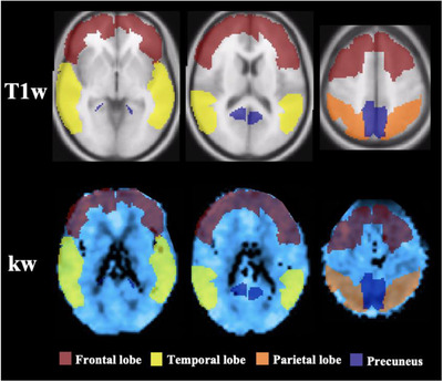
FIGURE 1.

Sample regions of interest (ROIs). Representations of masks used to extract values from the frontal lobe (maroon), temporal lobe (yellow), parietal lobe (orange), and precuneus (blue). ROIs are presented on a T1‐weighted image (first row) and on a representative participant's blood‐brain barrier water exchange (kw) map in normalized space (second row). Columns from left‐to‐right show horizontal slices moving in the inferior‐to‐superior direction. The same ROI masks were used for extraction of arterial transit time and cerebral blood flow values
