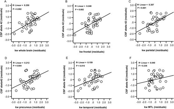
FIGURE 2.

Relationships between blood‐brain barrier water exchange (kw) values and cerebrospinal fluid (CSF) amyloid beta (Aβ)42 concentration. Scatter plots show kw values in the whole brain (A), frontal lobe (B), parietal lobe (C), precuneus (D), temporal lobe (E), and medial temporal lobe (F) plotted against CSF Aβ42 concentration. Plots show residual associations after controlling for age and sex
