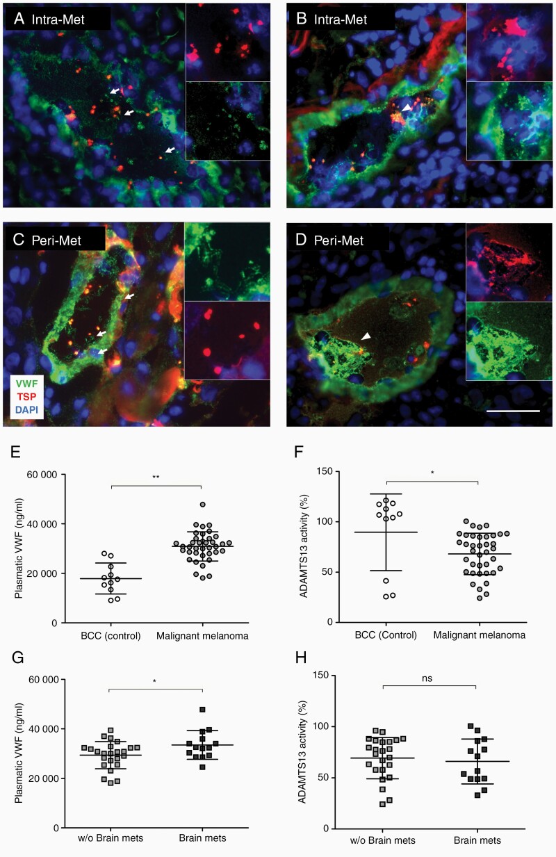
Figure 1.
Luminal VWF fibers and platelet aggregates are detected in the brain vessels of patients with BM. Immunofluorescence staining of VWF and platelet TSP shows luminal VWF fibers (arrows) and platelet aggregates (arrowheads) in Intra-Met (A, B) and Peri-Met (C, D) cerebral tissue from patients with BM (n = 7 patients). (E–H) VWF concentration and ADAMTS13 activity were measured in the plasma of BBC patients, used as control for non-metastatic skin tumor, and malignant melanoma patients with or without BM (n ≥ 11 patients/group). Ns = not significant, *P < .05, **P < .01, scale bar 50 μm.

