
Fig. 1|. Overview of structures of inactive Arp2/3 complex and activated Arp2/3 complex bound to actin filament pointed end and Dip1.
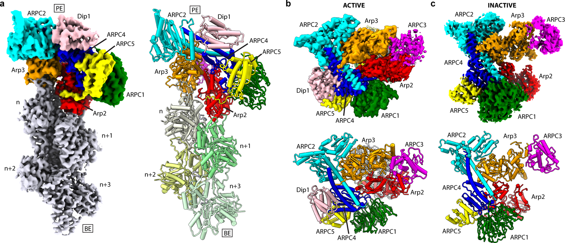
a, Reconstructed map (left) and atomic model (right) of actin filament pointed end with bound Arp2/3 complex and Dip1. PE: pointed end, BE: barbed end. b, Map (top) and atomic model (bottom) of activated Arp2/3 complex structure viewed from the pointed end looking down the helical axis of the nucleated actin filament. c, Map (top) and molecular model (bottom) of inactive Arp2/3 complex.