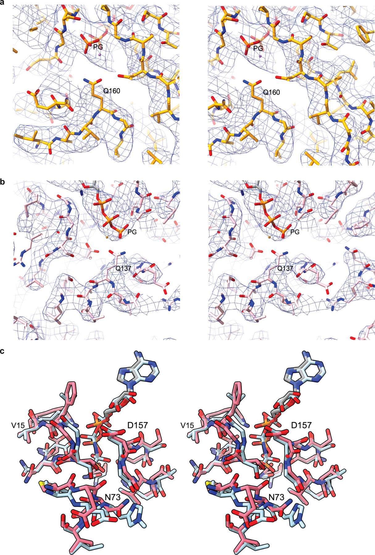
Extended Data Fig. 9|. Close up of nucleotide clefts of Arp2 and Arp3.

a, Stereo image of the nucleotide binding cleft of Arp3 showing the electron density of the modeled ATP phosphates and conserved catalytic residue Q160. b, Stereo image of the nucleotide binding cleft of Arp2 showing the electron density of the modeled ATP phosphates and conserved catalytic residue Q137. c, Stereo image of Arp2 from the activated structure (pink) superposed with actin (cyan) from the cryo-EM structure of AMP-PNP bound actin filaments15 showing the nucleotide and the nucleotide binding cleft.
