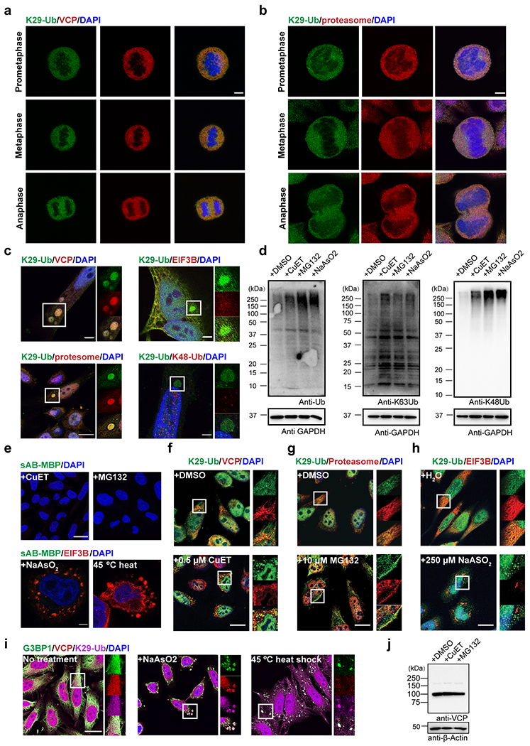
Extended Data Figure 5:

Image and gel panels in this figure are representative of two independent experiments; n = 2. a-b, Immunofluorescent staining of HeLa cells at prometaphase, metaphase, and anaphase of mitosis. Costaining for K29-Ub with VCP (panel a) or the proteasome (20S, panel b) is shown. Scale bars correspond to 5 μm. c, K29-linked ubiquitination is enriched in liquid droplets occasionally observed in normal HeLa cells. Costaining for K29-Ub with VCP, EIF3B, the proteasome (20S), and K48-Ub is shown. Scale bar in the bottom left panel corresponds to 20 μm; Scale bars in the other panels correspond to 5 μm. d, Western blots of total ubiquitin, K63- and K48-linked ubiquitin in HeLa cells treated with CuET (an inhibitor of the VCP cofactor Npl4, 1 μM for 4 h), MG132 (a proteasome inhibitor, 20 μM for 4 h), or sodium arsenite (an oxidative stress inducer, 500 μM for 1 h). e, Immunofluorescent staining of HeLa cells with sAB-MBP and costaining for EIF3B in HeLa cells treated with 500 μM sodium arsenite for 1 h or subjected to heat shock at 45 °C for 30 minutes. Scale bars in the top and bottom panels correspond to 20 and 5 μm, respectively. f-h, Immunofluorescent staining of HeLa cells treated with 0.5 μM CuET for 4 h, 10 μM MG132 for 4 h, or 250 μM sodium arsenite for 4 h. Costaining for K29-Ub with VCP, the proteasome (20S), and EIF3B is shown. Scale bars correspond to 20 μm. i, Immunofluorescent staining of HeLa cells treated with 500 μM sodium arsenite for 1 h or subjected to heat shock at 45 °C for 30 minutes. Costaining for K29-Ub with G3BP1 and VCP is shown. The scale bar corresponds to 20 μm. j, Western blot of VCP in HeLa cells treated with CuET (1 μM for 4 h) and MG132 (20 μM for 4 h).
