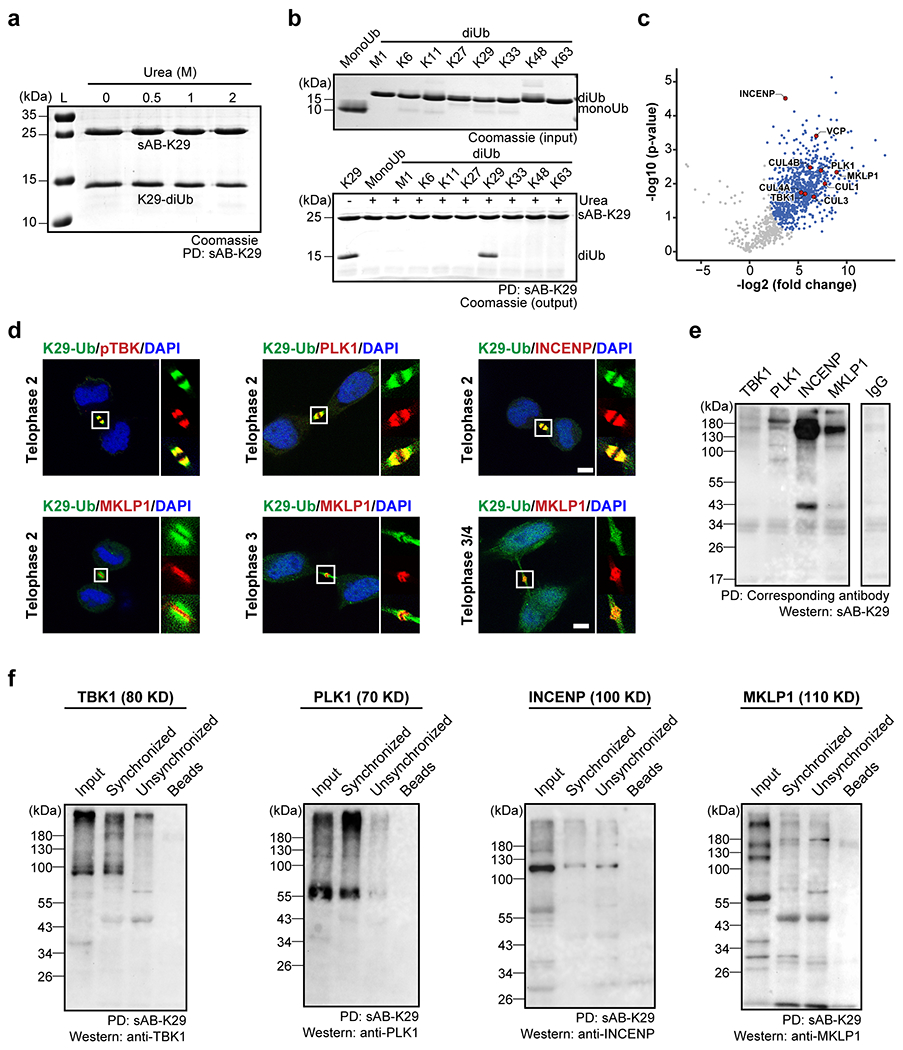
Extended Data Figure 10:

Gel panels and image panels in this figure are representative of two independent experiments; n = 2. a, sAB-K29 could be used to pull down K29-linked diUb in vitro under denaturing conditions (up to 2 M urea). b, sAB-K29 could be used to specifically pull down K29-linked diUb under denaturing conditions (2 M urea). c, Volcano plot of the quantitative mass spectrometry results after pull-down experiments in HeLa cells synchronized to the telophase of mitosis using sAB-K29 under denaturing conditions (1 M urea). Significant hits (FDR < 0.05) are colored blue, with those involved in midbody assembly highlighted in red. d, Immunofluorescent staining of HeLa cells at the telophase of mitosis. Costaining for K29-Ub with pTBK, PLK1, INCENP, and MKLP1 is shown. Scale bars correspond to 5 μm. e, Immunoprecipitation of synchronized HeLa cells under denaturing conditions using antibodies against TBK1, PLK1, INCENP, and MKLP1. Western blot was performed using sAB-K29 as the primary binder. Anti-IgG was used in the control experiment. f, Immunoprecipitation of HeLa cells (either synchronized to the telophase of mitosis or unsynchronized) using sAB-K29. Antibodies against TBK1, PLK1, INCENP, and MKLP1 were used for Western blot.
