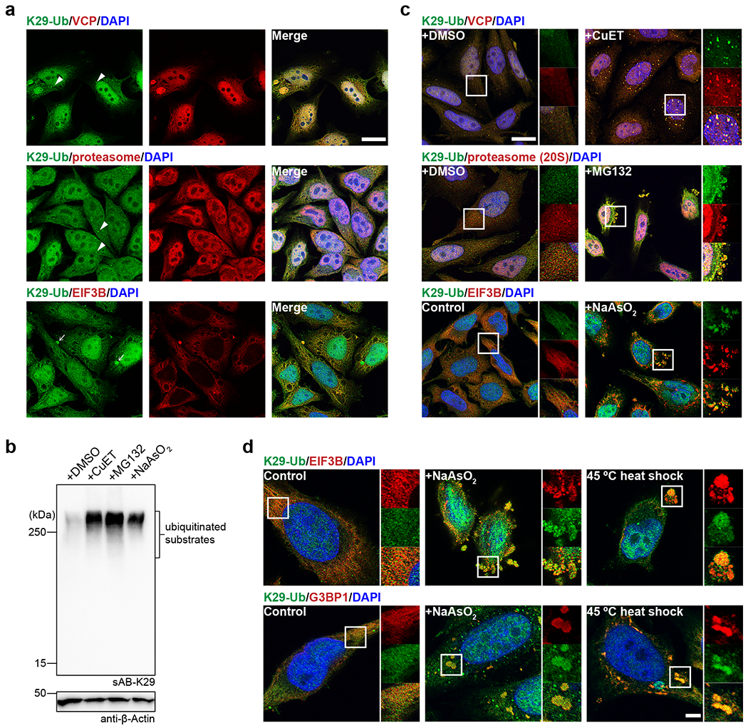
Fig. 4: K29-linked polyubiquitination is involved in protein homeostasis and the stress response.

Image panels and gel panels in this figure are representative of two independent experiments; n = 2. a, sAB-K29 was used for the immunofluorescent staining of untreated HeLa cells. Costaining for K29-Ub with VCP, the proteasome (20S), and EIF3B is shown. Arrowheads point to bright puncta stained by sAB-K29. Arrows point to liquid droplets stained by sAB-K29. b, K29-linked polyubiquitination was enhanced after HeLa cells were treated with CuET (an inhibitor of the VCP cofactor Npl4), MG132 (a proteasome inhibitor), or sodium arsenite (a stress response inducer). HeLa cells were treated with 1 μM CuET for 4 h, 20 μM MG132 for 4 h, or 500 μM NaAsO2 for 1 h, followed by Western blot analysis using sAB-K29 as the primary binder. c, Immunofluorescent staining of HeLa cells treated with 1 μM CuET for 4 h, 100 μM MG132 for 4 h, or 500 μM sodium arsenite for 1 h. Costaining for K29-Ub with VCP, the proteasome (20S), and EIF3B is shown. d, K29-linked polyubiquitination was enriched in stress granules. HeLa cells were treated with 500 μM sodium arsenite for 1 h or heat shocked at 45 °C for 30 minutes, followed by immunofluorescent staining. Costaining for K29-Ub with EIF3B and G3BP1 is shown. Scale bars in panels a and c: 20 μm, panel d: 5 μm.
