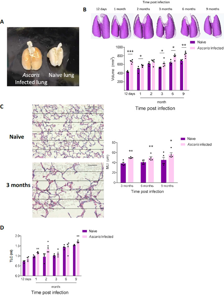
Fig 2. Ascaris suum induces chronic lung disease.
Mice were challenged with Ascaris suum at standard time intervals as previously described. (A) Gross appearance of lungs from age- and sex-matched Ascaris challenged mice and PBS naïve mice at day 12 post-infection (p.i.) demonstrating visibly larger lungs in the Ascaris challenged mouse (B) Representative 3-D reconstruction and post-expiratory aerated lung volume from Ascaris challenged mice and PBS naïve mice was calculated using micro computed tomography (microCT) at standard intervals p.i. and (C) Mean linear intercept (MLI) calculation from lung histology of mice p.i. compared to PBS naïve control mice at 3 months, 6 months and 9 months p.i.. Sections of lungs were specifically selected based on larval transition adjacent to bronchovascular bundles. (D) Lung compliance quantitated from mice at standard intervals p.i. measured as the total lung capacity (TLC) at a standard pressure (n≥5, mean±S.E.M, n.s.: not significant, *p<0.05, **p<0.01, ***p<0.001, using two tailed Student’s t-test. Magnification: 400×, Grid and scale bar: 50μm. Data are representative of at least two independent experiments).

