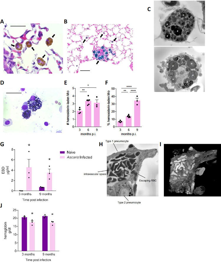
Fig 4. Ascaris induced vascular disruption and chronically induced hemosiderin-laden alveolar macrophages (MΦ).
Ascaris infected and PBS-challenged, naive mice were euthanized and lungs were harvested for histopathologic analysis at the indicated time interval. (A) Haematoxylin and eosin (H&E), and (B) Prussian blue staining of 5 μm lung sections. Arrow represents hemosiderin-laden macrophage. (C) Serial block electron microscopy (EM) image of hemosiderin-laden macrophages from lung sections demonstrates engulfed erythrocytes noted by the pound sign. These hemosiderin-laden macrophages were identified at 3 months, 6 months and 9 months post-infection (p.i.). (D) H&E staining of bronchoalveolar lavage fluid (BALF) cells demonstrates hemosiderin-laden macrophage that were also identified at 3 months, 6 months and 9 months p.i.. Absolute cell count (E) and percentage (F) of hemosiderin-laden macrophages from BALF at 3, 6 and 9 months p.i.. demonstrating the % of hemosiderin-laden macrophages increases chronically (G) Pulmonary extravasation of Evan’s blue dye (EBD) at 3 and 9 months p.i. to evaluate vascular integrity and leakage (H) EM image and (I) three-dimensional EM reconstruction of damaged pulmonary vascular structure demonstrating extravasation of the red blood cell (RBC) between a type-1 and type-2 pneumocyte moving from the endovasculature into the alveolar space due to edema. (J) Hemoglobin level from whole blood from infected mice at 3 and 9 months p.i. demonstrate chronic anemia in Ascaris infected mice. (Data are representative of at least two independent experiments)

