
Figure 4.
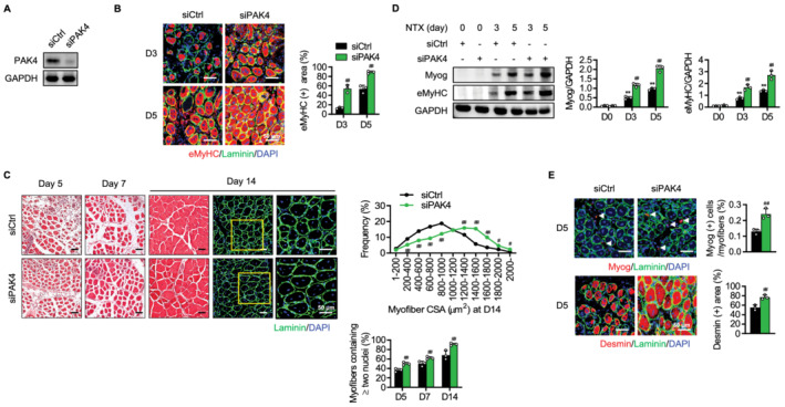
Acceleration of skeletal muscle regeneration in PAK4‐silenced mice. The tibialis anterior (TA) muscles of C57BL/6 mice were injected with scrambled siRNA (siCtrl) or siRNA against PAK4 (siPAK4) and then injured by intramuscular injection of NTX as shown in Figure 3A. (A) Western blotting analysis for PAK4 in PAK4 silenced muscles. (B) Immunofluorescence analysis of eMyHC‐positive fibres in TA muscles. (C) H&E and immunofluorescence analyses of sections. Average cross‐sectional area (CSA) of regenerating myofibres and the percentage of myofibres containing two or more centrally located nuclei per field were determined from immunofluorescence sections. (D) Time course analysis of myogenic markers by western blotting. (E) Immunofluorescence staining of Myog‐positive or desmin‐positive myofibres at Day 5. Arrowheads indicate MyoG‐positive myofibres. Values are mean ± SD. ** P < 0.01 vs. D0; ## P < 0.01 vs. siCtrl.