
Figure 4.
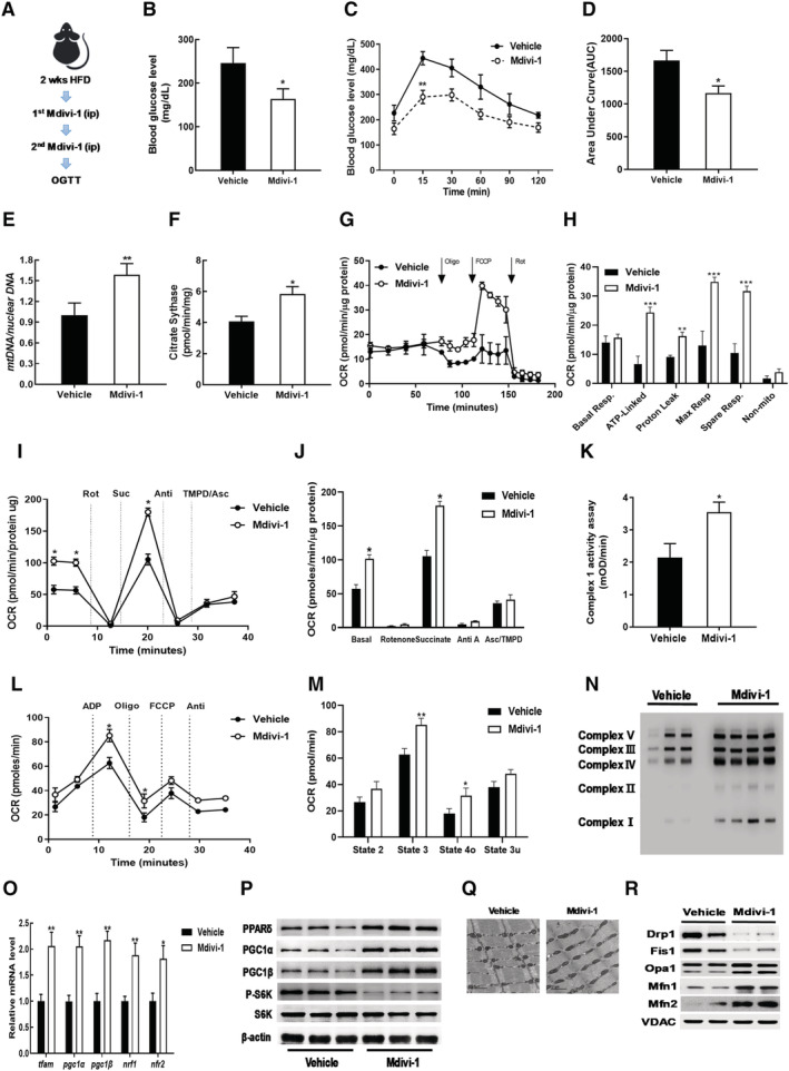
Mdivi‐1 administration improved acute IR via increased mitochondrial function. (A) Workflow for mdivi‐1 injections. (B) Blood glucose levels in plasma from vehicle DMSO or mdivi‐1 administrated HFD‐S mice. (C) Glucose tolerance test result. (D) AUC of glucose tolerance test result. (E) Mitochondrial DNA content in gastrocnemius muscles of mdivi‐1 treated mice versus normalized vehicle. (F) Citrate synthase activity in gastrocnemius muscles of mdivi‐1 group normalized to vehicle. (G) OCR measurements in EDL myofibres. (H) Analysis of mitochondrial respiration by OCR quantification. (I) Electron flow assay in isolated mitochondrial fraction of gastrocnemius muscles. (J) OCR quantification to measure complex‐dependent respiration. (K) Complex I activity in gastrocnemius muscles. (L) Mitochondrial respiratory coupling assay in isolated mitochondrial fraction of gastrocnemius muscles. (M) Quantification of respiratory coupling. (N) Expression of OXPHOS complex in gastrocnemius muscles. (O) Relative gene expressions for mitochondrial biogenesis and mtDNA replication measured by qRT‐PCR. (P) Western blot analysis for expression of PPARδ, PGC1α/β, S6K, and p‐S6K in gastrocnemius muscles. (Q) Mitochondrial morphology imaged by transmission electron microscopy (TEM). (R) Immunoblot analyses of proteins related to mitochondrial dynamics. Values expressed as means ± SEM and mean differences detected by Student's t‐test. *P < 0.05, **P < 0.01, ***P < 0.001 versus Vehicle, n = 5.