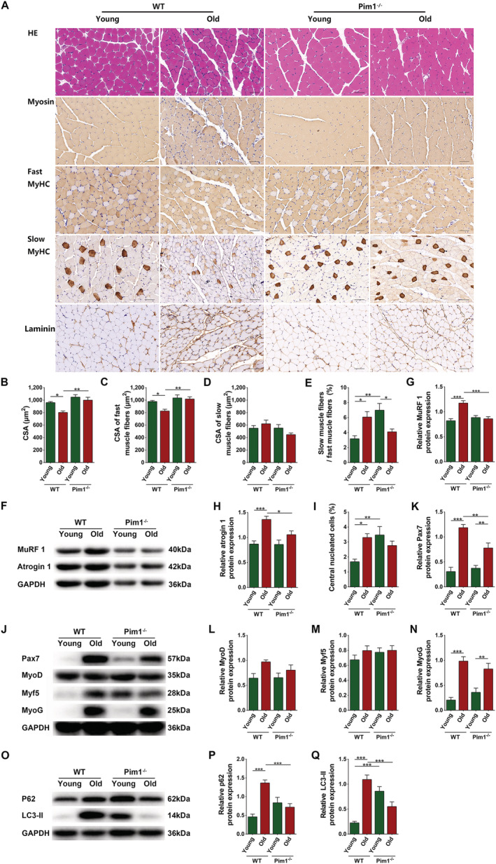
Figure 4.

Decreased intramuscular adipose tissue caused by Pim1 knockout alleviated skeletal muscle fibre atrophy and decreased the mobilization of satellite cells at the basal state in aging mice. (A) Haematoxylin–eosin (HE) staining and immunohistochemical staining of myosin, fast MyHC, slow MyHC, and laminin (scale = 50 μm). (B) Cross‐sectional area (CSA) of muscle fibres (μm2). (C) CSA of fast muscle fibres (μm2). (D) CSA of slow muscle fibres (μm2). (E) Ratio of the number of slow muscle fibres to the number of fast muscle fibres (%). (F) Detection of MuRF 1 and atrogin 1 expression through western blotting; GAPDH: internal reference. (G,H) Relative protein‐expression levels of MuRF 1 and atrogin 1. (I) Percentage of central nucleated muscle fibres in total muscle fibres (%). (J) Detection of Pax7, MyoD, Myf5, and MyoG expression through western blotting; GAPDH: internal reference. (K–N) Relative protein‐expression levels of Pax7, MyoD, Myf5, and MyoG. (O) Detection of P62 and LC3‐II expression through western blotting; GAPDH: internal reference. (P,Q) Relative protein‐expression levels of P62 and LC3‐II. N = 8; *P < 0.05, **P < 0.01, ***P < 0.001.
