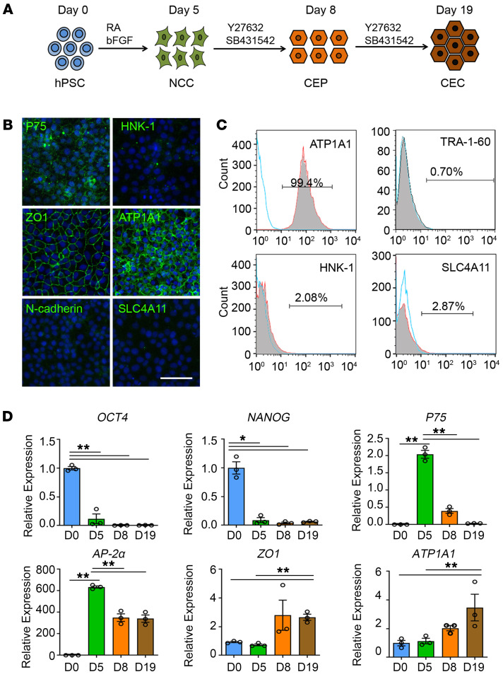
Figure 1. Induction and characterization of corneal endothelial precursors from hESCs.
(A) Schematic representation of the method used for the generation of NCCs, CEPs, and CECs. For the NCC induction, hPSCs are incubated in the neural crest differentiation medium with 4 ng/mL bFGF and 1 μM RA for 5 days. For the CEP and CEC induction, the medium is changed into corneal endothelial differentiation medium with 10 μM Y27632 and 1 μM SB431542 for a subsequent 3 days and 14 days. (B) Representative immunofluorescence staining of the neural crest markers P75 and HNK-1, and corneal endothelial markers ZO-1, ATP1A1, N-cadherin, and SLC4A11 in CEPs. Nuclei were stained with DAPI. Scale bar: 50 μm. (C) Flow cytometry analysis of ATP1A1, TRA-1-60, HNK-1, and SLC4A11 in CEPs. The experiments were repeated 3 times. (D) qPCR analysis of the gene expression during the 3 stages of hESC differentiation. n = 3, **P < 0.01, *P < 0.05 by 1-way ANOVA with Tukey’s HSD test.

