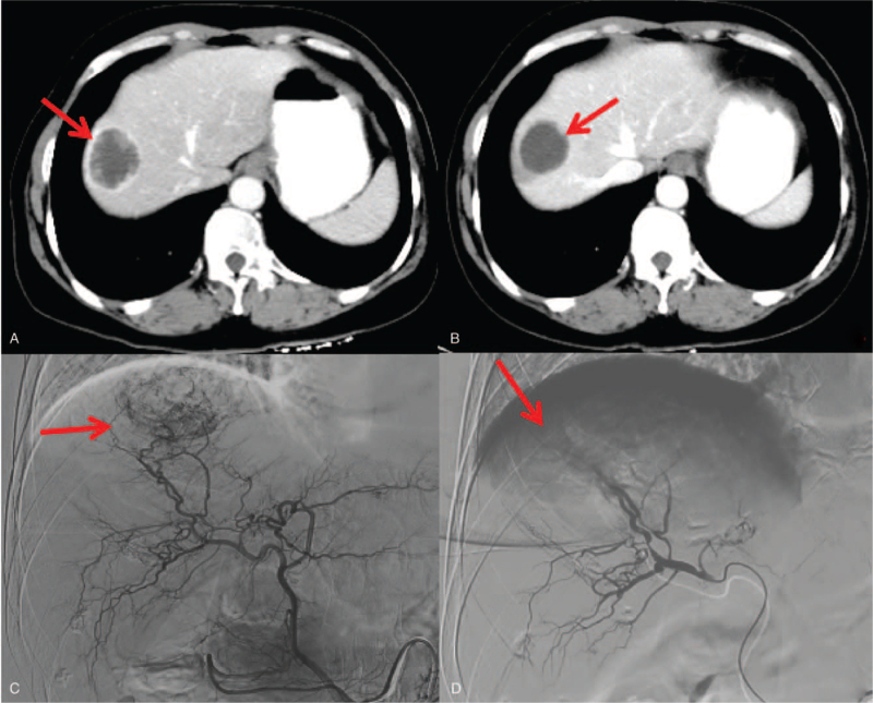
Figure 2.
Hepatic arteriography and post DEB-TACE treatment in a 47-year-old complete responder patient. A. Portal phase image of contrast-enhanced CT showed a low-attenuated lesion (arrow). B. At 3-months follow-up CT, the lesion decreased without contrast enhancement. C. Angiography during the DEB-TACE procedure revealed a comparatively hypervascular lesion. D. Shows the final hepatic angiogram. No remaining tumor blush is visible. CT = computed tomography, DEB-TACE = drug-eluting beads used for transarterial-chemoembolization.

