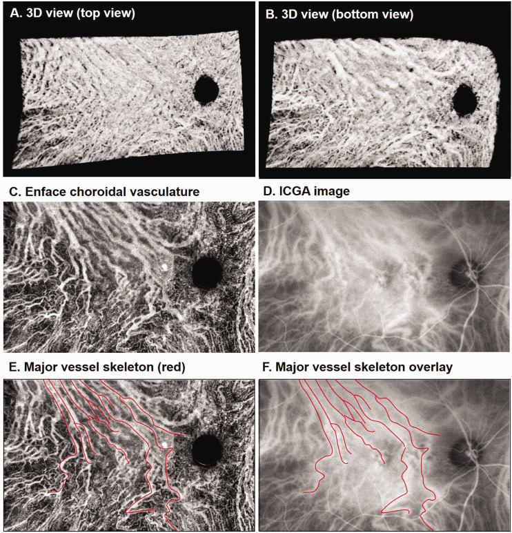
Figure 2.
Comparison of choroidal vasculature obtained from SS-OCT and ICGA. (a and b) 3D view of the choroid from a 75-year-old female patient with central serous chorioretinopathy. (c) En face images of choroidal vasculature generated via sum projection. (d) Cropped ICGA image at the same region. (e) Skeleton of major choroidal vessels (red) manually drawn on the en face image (c). (f) ICGA image with the vessel skeleton overlaid. (A color version of this figure is available in the online journal.)
ICGA: indocyanine green angiography.

