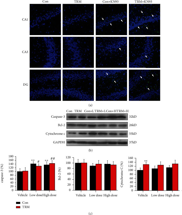
Figure 4.

CaMKII inhibition promotes apoptosis in the hippocampi of Wistar and TRM rats. (a) Images indicating TUNEL staining in the hippocampi (CA1, CA3, and DG regions) of Wistar rats, TRM rats, KN93-treated (high dose) Wistar rats, and KN93-treated (high dose) TRM rats. The arrows indicated apoptotic neurons. Scale bars: 100 μm. (b, c) The representative protein bands and data analysis of caspase-3, Bcl-2, and cytochrome c in the hippocampus of Wistar rats, TRM rats, KN93-treated (low dose) Wistar rats, KN93-treated (low dose) TRM rats, KN93-treated (high dose) Wistar rats, and KN93-treated (high dose) TRM rats. ∗p < 0.05, compared with the control Wistar group; #p < 0.05, compared with the control TRM group; ∗∗p < 0.01, compared with the control Wistar group; ##p < 0.01, compared with the control TRM group.
