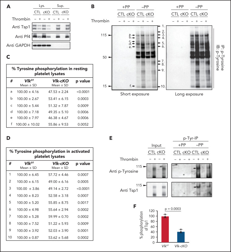
Figure 3.
Reduced tyrosine phosphorylation in resting and activated mouse platelets harboring deletion of Vlk compared with control. (A) Immunoblots of lysates (Lys.) and supernatants (Sup.) from Vlkf/f (CTL) and Vlk-cKO (cKO) platelets resting or stimulated with 5 U/mL thrombin for 15 minutes. Anti-Tsp1 and anti-PF4 antibodies were used as markers of platelet activation, and anti-GAPDH antibody was used to examine total protein levels in lysates. (B) Lysates from panel A were used to examine tyrosine phosphoproteins by immunoblot (IB) of p-Tyr–enriched fraction (IP). Black arrows indicate phosphobands decreased in Vlk-cKO (cKO) compared with Vlkf/f (CTL) in resting (a-f) and activated (1-10) platelets. Cumulative percent tyrosine phosphorylation in phosphorylated proteins indicated in representative figure in panel B from resting (C) and activated (D) control and Vlk-cKO lysates. Data are expressed as mean ± standard deviation (SD); n = 3 independent experiments. Quantifications were normalized to GAPDH protein expression. (E) Representative image of input and p-Tyr IP fraction from resting and thrombin-stimulated supernatants analyzed by using SDS-polyacrylamide gel electrophoresis (PAGE) with 8% gels and immunoblotting with anti–p-Tyr and anti-Tsp1 antibodies. Phenyl phosphate (PP) was used as negative control for nonspecific binding to p-Tyr beads. Black arrows indicate bands decreasing in cKO compared with CTL. (F) Cumulative percent phosphorylation of Tsp1 in activated supernatants from Vlk-cKO compared with CTL; n = 3 independent experiments.

