
Figure 3.
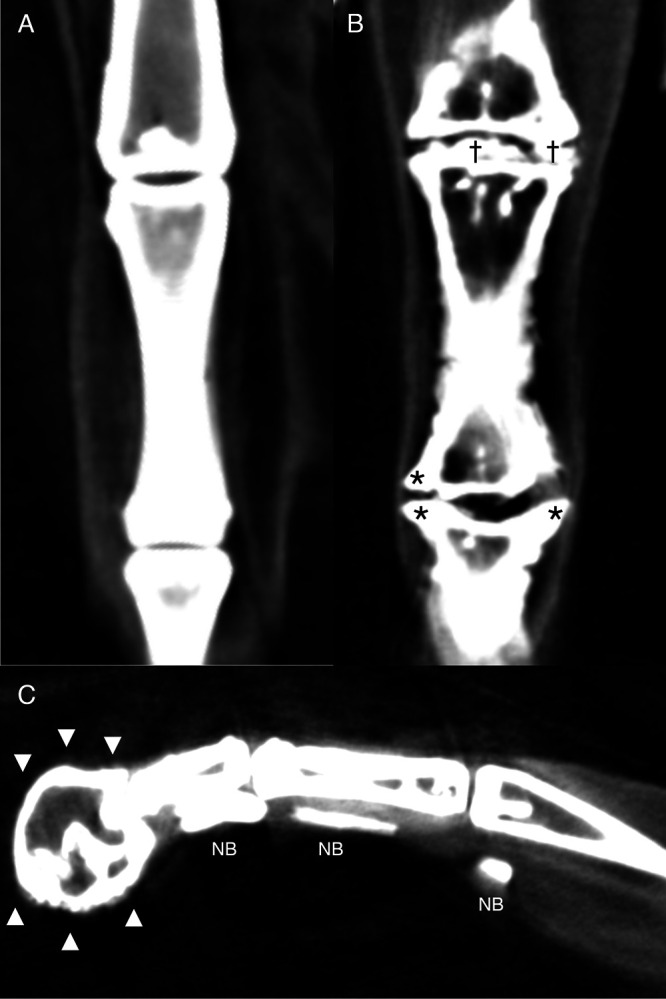
Reconstructed dorsal plane CT image of a normal MTP and IP joint (A), reconstructed dorsal plane CT image of a severely affected MTP joint and IP joint (B) with periarticular new bone formation (asterisks *) and new bone formation within the joint space (daggers †), and reconstructed sagittal plane CT image of a severely affected digit (C) with a misshaped distal phalanx surrounded by a rim of new bone (arrowheads xxx) and linear mineralization plantar to the digit within the extensor tendons (NB).