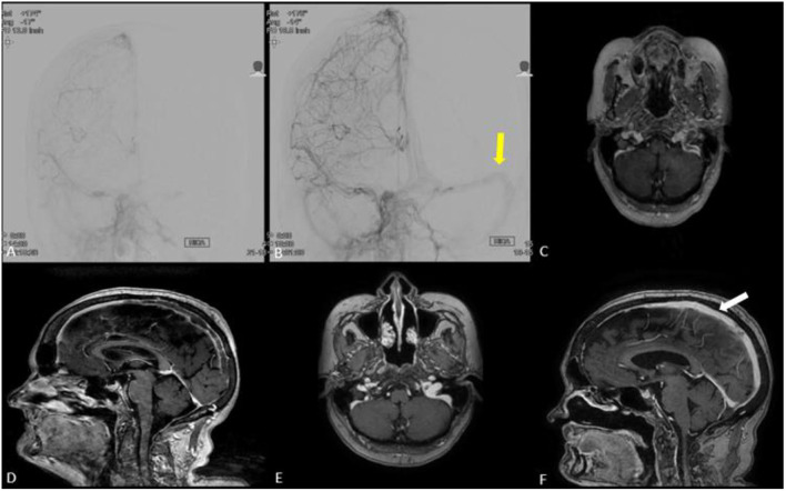
Figure 4.
(A,B) Anteroposterior cerebral angiogram run in (B) is post treatment demonstrating partial recanalization of posterior aspect of superior sagittal sinus and left transverse/sigmoid complex (yellow arrow) compared to pre-treatment (A). (C–F) MRI brain with and without contrast in axial (E) and coronal views (F) demonstrating near complete recanalization of superior sagittal sinus (white arrow) and left transverse/sigmoid complex at 3 months clinic follow-up compared to MRI brain obtained prior to venous thrombectomy (C,D).

