Figure 7.
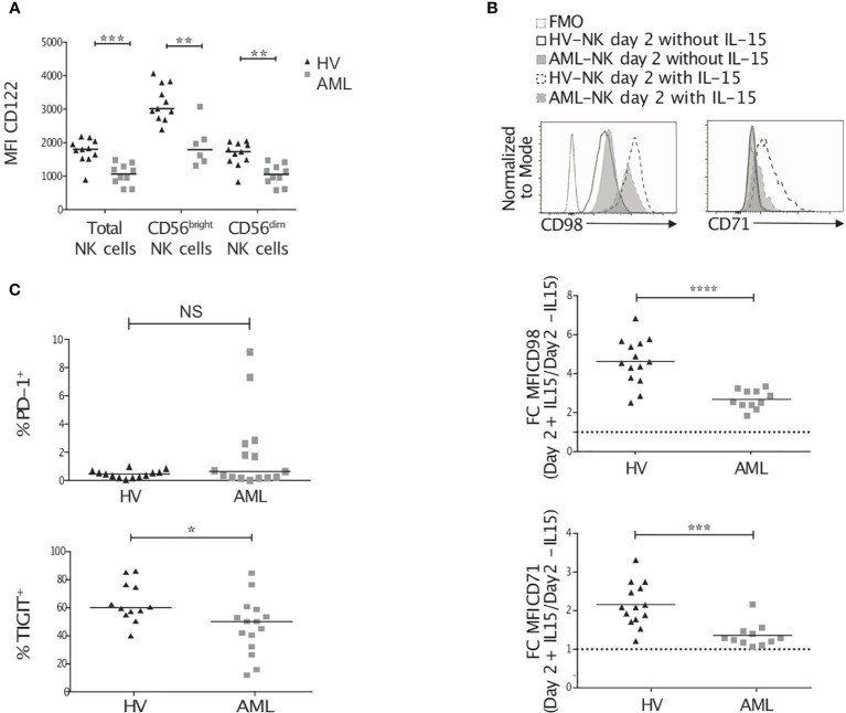
NK cells from AML patients have reduced IL-2/15Rβ and metabolic marker expression. Peripheral blood (PB) NK cells from healthy volunteers and AML patients were analyzed by flow cytometry for CD122 expression (A), CD98 and CD71 surface expression (B), and exhaustion marker expression (C). (A) Once thawed up, PB NK cells were stained for CD122, CD3, CD56 and CD45. Data show CD122 expression on CD45+/live total NK cells or CD56bright/dim NK cells. (B) Peripheral blood mononuclear cells were thawed and incubated for 48h in the presence or absence of 20ng/mL rhIL-15. The histograms show an example of CD98 and CD71 expression on NK cells at day 2 after culture with or without IL-15. The graphs show CD98 (top) and CD71 (bottom) expression ratio (stimulated versus unstimulated). (C) The level of expression of PD-1 (top) and TIGIT (bottom) in freshly thawed PB NK cells is depicted. Lines within each group correspond to the median. *p < 0.05, **p < 0.01, ***p < 0.001, ****p < 0.0001, as determined by Mann-Whitney test.

