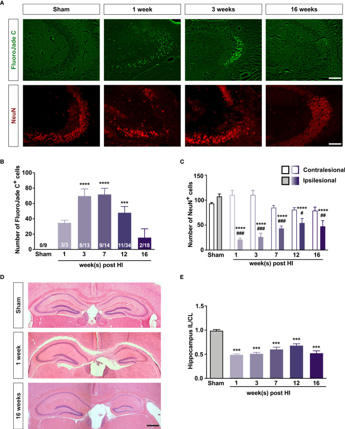
Figure 1.
Mild HI injury to developing brain leads to long-lasting neurodegeneration in the ipsilesional hippocampal CA3. (A) Representative images of ipsilesional CA3 in which degenerating neurons are visualized with FJC and all neurons are visualized with antibodies against NeuN 1–16 weeks after HI induction or 3 weeks after sham surgery. Scale bar = 50 µm. (B) Number of FJC+ cells and (C) number of NeuN+ cells in the CA3 of mice 1–16 weeks after neonatal HI induction or 3-7 weeks after sham surgery. (D) Representative images of hematoxylin-eosin–stained brain sections of mice 1 and 16 weeks after HI induction or 7 weeks after sham surgery. (E) Relative size of the ipsilesional hippocampus in mice 1–16 weeks after neonatal HI induction or 3–7 weeks after sham surgery. IL, ipsilesional; CL, contralesional. ***P < 0.001, ****P < 0.0005 vs sham; # P < 0.05, ## P < 0.005, ### P < 0.001 vs contralesional. Data were analyzed by one-way ANOVA and Dunnett’s posthoc test (B, E) or two-way ANOVA and Sidak’s posthoc test (C). Numbers in bars are number of positive mice/total number of mice (B). (C) n = 9 (sham); n = 3, 9, 14, 10, and 10 for 1, 3, 7, 12, and 16 weeks after HI induction, respectively. (E) n = 18 (sham); n = 3, 13, 14, 16, and 18 for 1, 3, 7, 12, and 16 weeks after HI induction, respectively. Values are mean ± SEM.

