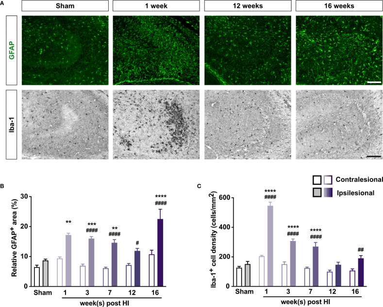
Figure 2.
Mild neonatal HI brain injury leads to long-lasting reactive gliosis. (A) Representative images of ipsilesional CA3 immunostained with antibody against GFAP to visualize astrocytes and with antibody against Iba-1 to visualize microglia 1–16 weeks after HI induction or 3 weeks after sham surgery. Scale bar = 50 µm. (B) Relative GFAP+ area and (C) density of Iba-1+ cells in CA3 of mice 1–16 weeks after HI induction or 3–7 weeks after sham surgery. IL, ipsilesional; CL, contralesional. **P < 0.005, ***P < 0.001, ****P < 0.0005 vs sham-operated mice; # P < 0.05, ## P < 0.005, ### P < 0.001, #### P < 0.0005 vs contralesional, by two-way ANOVA and Sidak’s posthoc test. n = 8 sham-operated mice; n = 3, 10–11, 12–13, 9, and 9 mice for 1, 3, 7, 12, and 16 weeks after HI induction, respectively. Values are mean ± SEM.

