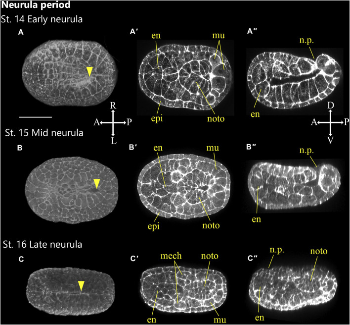
FIGURE 5.
Ascidiella aspersa embryos at neurula period. Embryos are stained with Alexa Fluor™ 546 Phalloidin. Three-dimensional reconstructed images at (A) Stage 14 (early neurula), (B) Stage 15 (mid neurula), and (C) Stage 16 (late neurula). Three-dimensional reconstructed images at (A) Stage 14 (early neurula), (B) Stage 15 (mid neurula), and (C) Stage 16 (late neurula). (A–C) show the dorsal views of the embryos. (A′–C′) show the cross-section, and (A′′–C′′) show longitudinal section of (A–C), respectively. Embryo direction is shown by A, anterior; P, posterior; D, dorsal, and V, ventral. Arrowheads indicates the anterior edge of neural tube closure. en, endoderm; epi, epidermis; mech, mesenchyme; mu, muscle; noto, notochord; n.p., neuropore. Scale bar: 50 μm.

