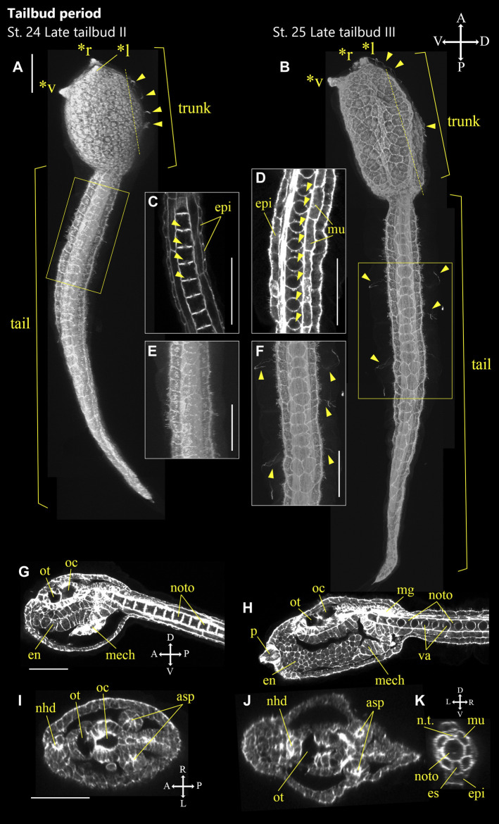
FIGURE 7.
Ascidiella aspersa embryos at the latter of tailbud period. Embryos were stained with Alexa Fluor™ 54 Phalloidin. Three-dimensional reconstructed images at (A) Stage 24 (late tailbud II) and (B) Stage 25 (late tailbud III). (A,B) show the lateral views of the embryos. (C,D) Enlarged longitudinal section of the area inside the yellow frame in (A,B), respectively. Arrowheads indicate vacuoles in notochord cells. (E,F) Enlarged image of the area inside the yellow frame in (A,B), respectively. (G,H) Longitudinal section of (A,B), respectively. (I,J) Cross-section of the trunk. The positions of the sections are indicated by dashed lines in (A,B). (K) Cross-section of the tail in (B). Embryo direction is shown by A, anterior; P, posterior; D, dorsal; V, ventral. In (A,B, F), arrowheads indicate the cilia of epidermal sensory neurons. Asterisks indicates the papillae by *l: left papilla, *r: right papilla, *v: ventral papilla. asp, atrial siphon primordium; en, endoderm; epi, epidermis; es, endodermal strand; mech: mesenchyme; mg, motor ganglion; mu, muscle; nhd, neurohypophyseal duct; noto, notochord; n.t., neural tube; oc, ocellus; ot, otolith; p, papilla; and va, vacuole. Scale bar: 50 μm.

